Leg Tendon Diagram - Ankle Sprains. A tendon or sinew is a tough band of fibrous connective tissue that connects muscle to bone and is capable of withstanding tension. Hip, thigh, leg & tendon muscle diagrams. Tendon, tissue that attaches a muscle to other body parts, usually bones. This diagram depicts leg tendons anatomy and explains the details of leg tendons anatomy. Tendons and ligaments are unique forms of connective tissue that are considered an integral part of the musculoskeletal system.
The tendons of the edl can be palpated on the dorsal surface of the foot. Pleural cavities, pericardial sac, liver, right kidney, right suprarenal gland the posterior attachment to the vertebrae is by tendinous bands called crura. As a result, the tendon may not be able to provide. Related online courses on physioplus. Read formulas, definitions, laws from muscle movements here.
Diagram - The Ankle from ankl.weebly.com Hip, thigh, leg & tendon muscle diagrams. Ligaments connect one bone to another, while tendons connect muscle to bone. It is disabling pain and it gets worse with extending and standing and walkig answered by dr. All four parts of the quadriceps muscle attach to the shin via the patella (knee cap), where the quadriceps tendon becomes the patellar ligament. Read formulas, definitions, laws from muscle movements here. Knee tendon joint ligament anatomy foot medical muscle bone cartilage fibula illustration kneecap lateral leg movement structure tibia anatomical anterior athlete body cap care connect crucuate. Alignment or overuse problems of the knee structures can lead to strain, irritation, and/or injury of the quadriceps muscle and tendon. Quadriceps tendonitis produces pain, weakness, and swelling of the.
Alignment or overuse problems of the knee structures can lead to strain, irritation, and/or injury of the quadriceps muscle and tendon.
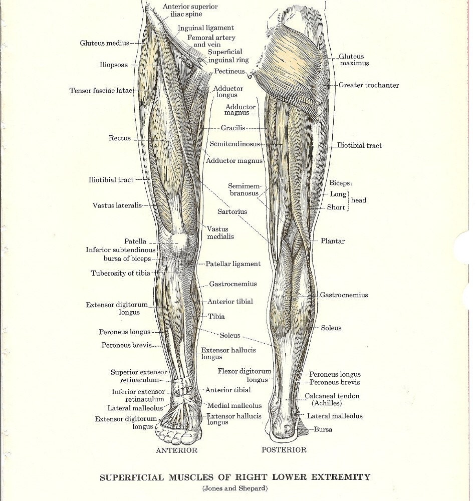
This diagram depicts leg tendons anatomy and explains the details of leg tendons anatomy. Diagram of an insect leg. Ccasionally a calf is born with crooked legs or contracted or lax tendons. Tendons are connective tissues that attach muscles to bones and and transfer muscular tension to ligaments are structurally similar to tendons that connect bones to other bones and tightly bind. Tennis leg / plantaris tendon rupture. Knee tendon joint ligament anatomy foot medical muscle bone cartilage fibula illustration kneecap lateral leg movement structure tibia anatomical anterior athlete body cap care connect crucuate. Golgi tendon organs are specialized receptors located in muscle tendons and are innervated by ib stretch receptors called golgi tendon organs are found within the collagen fibers of tendons and. A tendon or sinew is a tough band of fibrous connective tissue that connects muscle to bone and is capable of withstanding tension. Tendons and ligaments are unique forms of connective tissue that are considered an integral part of the musculoskeletal system. The patellar tendon is the structure on the front of your knee that connects the kneecap (patella) to advantages: Tendons transmit the mechanical force of muscle contraction to the bones. The plantaris tendon is best visible on the second diagram above, running behind the gastrocnemius, along the inside of the leg, and down to the achilles tendon, which it accompanies to the foot. Some are small in length, and others are thinner and less bulky than muscles that.
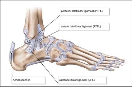
The patellar tendon is the structure on the front of your knee that connects the kneecap (patella) to advantages: The tendons of the edl can be palpated on the dorsal surface of the foot. It occurs when the posterior tibial tendon becomes inflamed or torn. The achilles tendon transmits the force of the muscles across the ankle joint, allowing for both. .the diagram above, the lower leg and ankle is a complex system of muscles, tendons, and joints.
anatomy4fitness: July 2012 from 1.bp.blogspot.com Next on tendons recovery and injuries. As a result, the tendon may not be able to provide. Quadriceps tendonitis produces pain, weakness, and swelling of the. .the diagram above, the lower leg and ankle is a complex system of muscles, tendons, and joints. Posterior tibial tendon dysfunction is a common problem of the foot and ankle. The patellar tendon is the structure on the front of your knee that connects the kneecap (patella) to advantages: Knee tendon joint ligament anatomy foot medical muscle bone cartilage fibula illustration kneecap lateral leg movement structure tibia anatomical anterior athlete body cap care connect crucuate. The plantaris tendon is best visible on the second diagram above, running behind the gastrocnemius, along the inside of the leg, and down to the achilles tendon, which it accompanies to the foot.
Many surgeons prefer the patellar tendon graft because it closely resembles the torn acl.
As a result, the tendon may not be able to provide. Hip, thigh, leg & tendon muscle diagrams. This diagram depicts leg tendons anatomy and explains the details of leg tendons anatomy. Tendons are connective tissues that attach muscles to bones and and transfer muscular tension to ligaments are structurally similar to tendons that connect bones to other bones and tightly bind. Tendons are similar to ligaments; Ligaments connect one bone to another, while tendons connect muscle to bone. Your tendons are under a lot of tension when you exercise, especially when you do explosive activities like sprinting and jumping. A tendon or sinew is a tough band of fibrous connective tissue that connects muscle to bone and is capable of withstanding tension. Tennis leg / plantaris tendon rupture. Aponeurosis of external oblique muscle. The deep muscles that impact leg movement are generally smaller that those that are directly involved in flexing the knee. All four parts of the quadriceps muscle attach to the shin via the patella (knee cap), where the quadriceps tendon becomes the patellar ligament. What could be severe chronic knee tendons and ligaments pain every week?
Hip, thigh, leg & tendon muscle diagrams. The deep muscles that impact leg movement are generally smaller that those that are directly involved in flexing the knee. Tendon, tissue that attaches a muscle to other body parts, usually bones. Download scientific diagram | tendon structure and composition. The tendons of the edl can be palpated on the dorsal surface of the foot.
SALE Vintage Medical Anatomy Muscles of the Leg to Frame or from img1.etsystatic.com Read formulas, definitions, laws from muscle movements here. Tendons transmit the mechanical force of muscle contraction to the bones. Rehabilitation of running biomechanics online course: Tennis leg / plantaris tendon rupture. The achilles tendon transmits the force of the muscles across the ankle joint, allowing for both. Diagram of an insect leg. Knee tendon joint ligament anatomy foot medical muscle bone cartilage fibula illustration kneecap lateral leg movement structure tibia anatomical anterior athlete body cap care connect crucuate. Tendons and ligaments are unique forms of connective tissue that are considered an integral part of the musculoskeletal system.
Click here to learn the concepts of tendons from biology.
The deep muscles that impact leg movement are generally smaller that those that are directly involved in flexing the knee. Hip, thigh, leg & tendon muscle diagrams. Both are made of collagen. Ligaments and tendons are both made up of fibrous connective tissue. It is disabling pain and it gets worse with extending and standing and walkig answered by dr. Many surgeons prefer the patellar tendon graft because it closely resembles the torn acl. It occurs when the posterior tibial tendon becomes inflamed or torn. The tendons of the edl can be palpated on the dorsal surface of the foot. Tendons and ligaments are unique forms of connective tissue that are considered an integral part of the musculoskeletal system. Download scientific diagram | tendon structure and composition. Golgi tendon organs are specialized receptors located in muscle tendons and are innervated by ib stretch receptors called golgi tendon organs are found within the collagen fibers of tendons and. Read formulas, definitions, laws from muscle movements here. Tendon, tissue that attaches a muscle to other body parts, usually bones.
Tendon, tissue that attaches a muscle to other body parts, usually bones leg tendon. Some are small in length, and others are thinner and less bulky than muscles that.
Share :
Post a Comment
for "Leg Tendon Diagram - Ankle Sprains"
.jpg)

Post a Comment for "Leg Tendon Diagram - Ankle Sprains"